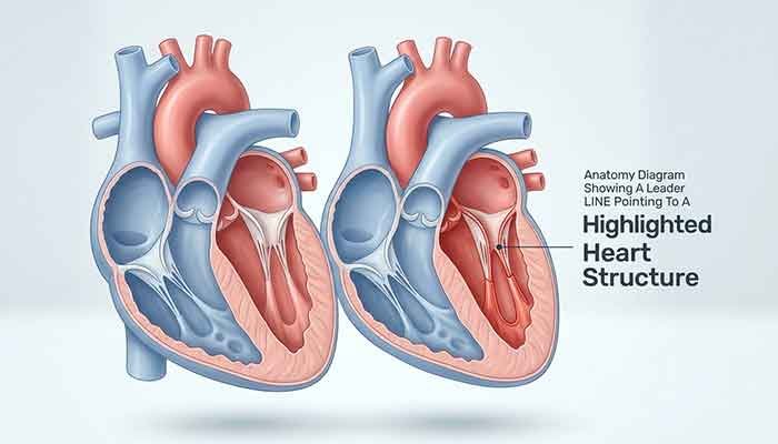
I still remember sitting in my dorm room at 11 PM, staring at a McGraw Hill Anatomy & Physiology Revealed screenshot, with a little arrow pointing at some unlabeled blob on a heart diagram. The question just said: “Which structure is highlighted and indicated by the leader line?” And I had absolutely no idea what I was looking at. Sound familiar?
That moment is exactly why I’m writing this. As someone who’s spent way too many late nights messing around with anatomy software, diagram tools, and quiz platforms (yeah, I’m that guy who nerds out over UI details most people ignore), I’ve learned that leader line questions trip people up not because the anatomy is impossible, but because nobody actually explains how to read a leader line in the first place. So let’s fix that today.
What Is A Leader Line In Anatomy Diagrams?
A leader line is simply the thin line — sometimes straight, sometimes slightly curved — that connects a label, a highlighted region, or a question marker to a specific structure on an anatomical image. Think of it like a tiny signpost saying “hey, I’m talking about THIS exact spot, not the general area around it.”
Here’s the part that confused me at first: leader lines don’t just point at things randomly. They usually terminate at one precise, tiny point on the structure. If the line’s tip lands even slightly off, on a border, or between two tissues, you can misidentify the structure completely. That precision is the whole point of the question — it’s testing whether you can distinguish a structure from its neighbors, not just recognize the general region of the body.
How Do You Identify A Highlighted Structure In An Anatomical Diagram?
Quick summary: To identify a highlighted structure, trace the leader line back to its exact endpoint, note the surrounding tissue type and position (superior/inferior, proximal/distal), and cross-reference with the diagram’s system (skeletal, cardiovascular, integumentary, etc.) before selecting an answer.
Here’s my personal process, refined after way too many wrong guesses:
- Zoom in first. Most platforms, including McGraw Hill APR, let you zoom. Do it every single time.
- Follow the line to its tip, not its middle. The tip is the actual target.
- Check the layer. Is it superficial (skin), deep (organ), or somewhere in between (fascia, muscle)?
- Use the system context. A heart diagram won’t highlight a skin gland, obviously — but it might highlight a vessel that looks like a chamber at first glance.
- Compare color and texture if the software renders realistic tissue — arteries, veins, and nerves often have subtly different shading.
Read More: Is Ziddu Safe? My Comprehensive Ziddu.com Review
Which Structure Is Highlighted And Indicated By The Leader Line — McGraw Hill APR
This is probably the most searched version of this question, and I get why. McGraw Hill’s Anatomy & Physiology Revealed (APR) is used in a ton of college A&P courses, and its quiz mode loves throwing leader line questions at students without much warning.
Here’s the honest truth, though: I can’t tell you the exact structure for your specific screenshot without seeing it, because APR pulls from dozens of different cadaver images, models, and rotation angles. Anyone online claiming there’s one universal answer to “which structure is highlighted and indicated by the leader line apr” is oversimplifying it. What I can do is walk you through the most commonly tested structures so you can recognize them fast.
Read More: Almawadahit .com Blog Review: Features & Safety
Common APR Leader Line Targets by System
| System | Frequently Highlighted Structures | What Makes It Tricky |
|---|---|---|
| Cardiovascular | Superior Vena Cava, Aortic Arch, Coronary Sinus | Vessels look similar in cross-section |
| Nervous | Medulla Oblongata, Corpus Callosum, Thalamus | Brain regions blend without clear borders |
| Integumentary | Eccrine Sweat Gland, Hair Follicle, Sebaceous Gland | Tiny structures crammed close together |
| Skeletal | Foramen Magnum, Zygomatic Arch, Styloid Process | Bone landmarks look alike from certain angles |
| Muscular | Origin vs. Insertion points | Leader line often sits right on the tendon boundary |
Honestly, the cardiovascular and integumentary sections give students the most grief — I know they got me. The Superior Vena Cava especially gets confused with the aorta way more than it should, mostly because of how flat 2D renderings can be.
My Personal Experience With Leader Line Anatomy Questions
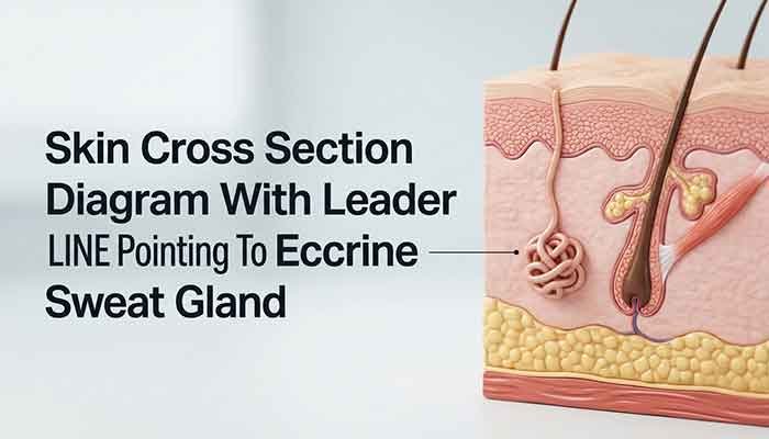
Okay, story time. A few semesters back, I was helping my cousin study for her A&P practical using APR, and we hit a skin cross-section slide with a leader line pointing at what looked like a tiny coiled tube buried in the dermis. She guessed “hair follicle” instantly — reasonable guess, honestly. But when I zoomed in (way more than she had), the coiled structure was actually an Eccrine Sweat Gland, not a follicle. The follicle was right next to it, angled diagonally, and its base was what was throwing her off.
What I learned from that moment, and honestly kept using since, is that leader line questions are less about “do you know anatomy” and more about “can you slow down and actually look.” She knew the material. She just moved too fast. Once I told her to always zoom before answering, her accuracy on those specific question types jumped noticeably in the next practice set. That one small habit change made a bigger difference than any amount of re-reading the textbook chapter did.
I’ll admit, I was a little smug about catching that gland/follicle mix-up. But it genuinely changed how I approach any labeled diagram now, whether it’s anatomy, a circuit board schematic, or a UI wireframe at work. Precision over speed, every time.
What Brain Structure Is Highlighted And Indicated By The Leader Line?
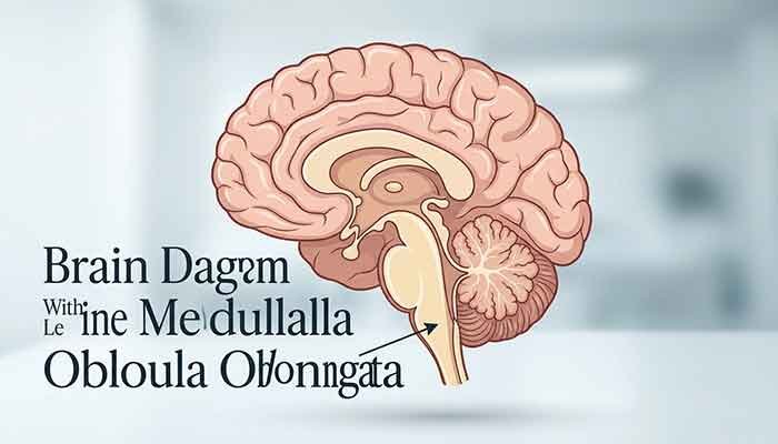
Quick summary: In most brain diagrams, leader lines commonly target the Medulla Oblongata, Corpus Callosum, Thalamus, or Cerebellum, since these regions are frequently tested for their distinct location and function relative to neighboring structures.
Brain diagrams are rough because there’s no hard line between a lot of regions — it’s more of a gradient. A few tips that helped me:
- The Medulla Oblongata sits at the very base, connecting to the spinal cord — if the leader line points low and central, that’s your first guess.
- The Corpus Callosum is the thick white band connecting the two hemispheres — usually shown mid-sagittal.
- The Thalamus sits centrally, just above the brainstem, often mistaken for the hypothalamus right below it.
What Cardiovascular Structure Is Highlighted And Indicated By The Leader Line?
Quick summary: Cardiovascular leader line questions frequently target major vessels like the Superior Vena Cava, Aorta, or Pulmonary Artery, since these structures sit close together and require careful attention to size, position, and curvature to tell apart.
If the leader line hits a large vessel near the top-right of the heart, entering the right atrium, that’s almost always the Superior Vena Cava. If it’s arching over the top like a candy cane, that’s the Aortic Arch. Vessel curvature is honestly your best friend here.
Which Layer Of Skin Is Highlighted And Indicated By The Leader Line?
Quick summary: Skin diagrams typically highlight the epidermis, dermis, or hypodermis, along with embedded structures like the eccrine sweat gland or hair follicle, and the leader line’s depth in the cross-section tells you which layer or structure is targeted.
Depth is everything here:
- Epidermis — thin, outermost, no blood vessels visible
- Dermis — thicker, contains glands, follicles, and vessels
- Hypodermis — fatty layer, deepest, loosely connected tissue
How To Solve McGraw Hill Anatomy APR Leader Line Questions (Step-By-Step)
- Screenshot and zoom — don’t answer directly off the small preview window.
- Identify the system — cardiovascular, integumentary, skeletal, etc.
- Note the exact tip location of the leader line, not the general area.
- Eliminate obviously wrong answers based on tissue type or position.
- Cross-check with your textbook diagram for that same region before finalizing.
Does this take longer than just guessing? Yeah, a bit. But it genuinely saves you from the frustrating “so close but wrong” answers that leader line questions are notorious for.
FAQ
Q: Why do leader line questions feel harder than regular label questions?
Because there’s no text label to lean on — you have to rely purely on position, depth, and shape, which forces you to actually understand the anatomy instead of memorizing a word next to a picture.
Q: Is there one universal answer to “which structure is highlighted and indicated by the leader line”?
No, and I want to be upfront about that. The answer always depends on the specific diagram, rotation, and system shown in your exact question. Anyone claiming a single fixed answer for every version of this question isn’t being fully accurate.
Q: What’s the fastest way to get better at these questions?
Honestly, repetition with zoomed-in review. Go slow the first few times, and speed naturally follows once the shapes become familiar.
Q: Do leader line questions show up outside of McGraw Hill APR?
Yes — plenty of anatomy quizlet sets, textbook practicals, and even some skeletal and dental diagrams use the same leader line format.
Q: Can two students get the same leader line question with different correct answers?
Sometimes, yes, if the software randomizes which diagram or rotation appears. That’s part of why I always recommend confirming against your own course’s specific image rather than a generic online answer key.
Martin’s Final Tip
If there’s one thing I want you to walk away with, it’s this: slow down before you answer. Leader line questions aren’t designed to trick you — they’re designed to reward the person who actually looks closely instead of pattern-matching from memory. Zoom in, trace that line to its exact tip, and think about the layer and system before you commit to an answer.
Have you run into a leader line question that completely stumped you? I’d genuinely love to hear which structure got you — drop it in the comments, and let’s compare notes on the tricky ones.